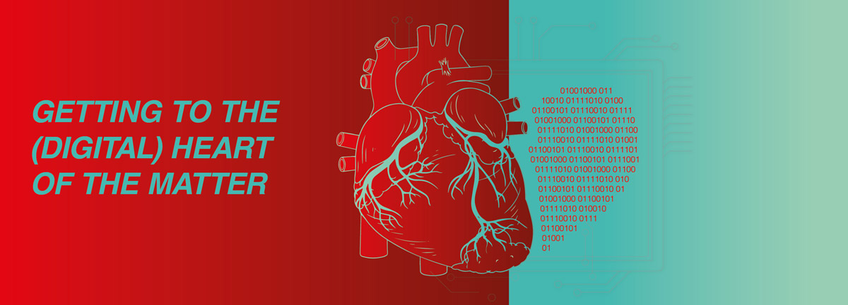
Digital twins – getting to the heart of the matter
The patient is experiencing auricular fibrillation. Her medication is having only a limited effect. Could medical intervention help or would the team of doctors be better to hold off? What types of treatment are most likely to help her recover? Decisions such as these are part and parcel of daily life working in cardiology. In most cases, however, they are decisions based on standards, empirical values and have very little to do with the specific condition of the individual patient’s heart.
Graz Medical University is developing a technology that should make treatment decisions such as these more precise and more tailored to the individual: A digital heart twin, an accurate replica of the organ, should reveal how one specific heart would react to different methods of intervention. The virtual heart model is designed to help the hospital clinicians on a daily basis to determine the most effective and safest treatment option for this individual patient and other patients.
The first step towards personalised cardiology
Every human heart is unique – in its size, shape and even the exact way it beats and functions. By the same token, every heart is unique in how it becomes ill and how it heals. For instance, for around 30 % of all patients, standardised pacemaker treatment is not effective. Conventional treatment methods for cardiac arrhythmia are still all too often based on average values and rarely call upon detailed medical reports.
This is precisely where the idea of the digital heart twin comes into play: It realistically portrays the heart of a specific patient. As such, the virtual model allows for a more accurate diagnosis and, in turn, personalised treatment strategies.
Research group in Graz has been developing digital heart twins for years
A research team based at Graz Medical University and Graz Technical University, led by heart modelling expert Gernot Plank, has already been working for several years on the development of digital twins of human hearts. The original idea has since evolved into a model that is essentially ready to go: At Graz Hospital, anatomically correct digital twins are already being produced. It is now a matter of fine-tuning the technology, until it can be routinely used before and during an operation.
After all, creating a highly accurate replica of a heart with its three-dimensional geometry and the many mechanical, physical and electrochemical processes it performs is a mammoth digital task.
Extensive modelling of the heart’s geometry and functionality
Even creating a generic heart model is highly complex. Modelling requires special mathematical processes, algorithms, hardware, software and enormous processing power to complete billions of calculations every second.
The composition of a virtual heart twin therefore begins by collecting patient imaging data through MRI and CT scans. Using this data, together with segmentation algorithms, the patient-specific heart structures of the atria, heart chambers and valves are digitally reconstructed. For this step, the researchers are increasingly turning to machine learning. The geometric data provided by CT and MRI scans, including pathological findings, are then combined with functional information from the ECG, such as heart rate, rhythm and propagation of heart pulses. Only when anatomical and functional parameters are linked can a realistic digital heart model be made.
The electrical propagation in the heart is fundamental for the simulation
The simulation of the heartbeat plays a central role in the development of the digital heart twin. The heartbeat is triggered by an electrical wave that propagates through the heart muscle tissue – from sinus nodes via the conduction system to the muscle cells of the atria and heart chambers.
The propagation of the electrical waves that are responsible for contracting the heart is simulated in the digital twin by means of eikonal equation, which calculates the time of stimulation at every point of the tissue. For this, the researchers had to consider that the electrical wave does not propagate everywhere at the same speed. For instance, the speed reached along muscle fibres is greater than the speed reached crossing them. Other decisive factors include the individual shape and wall structure of the heart, as well as pathological changes, such as scarring or fibrosis. These can delay or even stop the propagation of the pulse.
This precise reproduction of electrical heart activity underpins patient-specific treatment, because it can predict, for example, the impact of any planned procedure.
The future of digital heart twins – balancing precision with responsibility
Digital heart twins open up new pathways for cardiology, by facilitating more accurate, personalised and predictive diagnoses and treatment plans in cases of cardiac arrhythmia. In future, this technology should enable doctors to play through various treatment options on the computer model before making a final decision on any procedure – with no risk to patients.
At the same time, this source of enormous potential still faces a few technical, data protection and bureaucratic hurdles. For instance, there is the elaborate modelling, which requires high levels of processing and has to be approved for everyday clinical use before it can be used extensively. There are also the ethical questions, as encountered by other medical applications of AI-assisted diagnostics: Who makes the ultimate decisions regarding the simulated heart? Who is responsible for treatment decisions, if an AI system is involved in the modelling?
For daily work on major objectives and visions:
Carl ROTH can supply you with all manner of aids to facilitate your research and laboratory work. To see what we have to offer, please visit our online shop: www.carlroth.com
Sources:
https://www.medunigraz.at/news/detail/digitale-zwillinge-des-herzens
https://www.tugraz.at/news/artikel/wie-simuliert-man-den-digitalen-zwilling-des-herzens
https://www.sn.at/panorama/wissen/digitales-herzmodell-graz-therapien-183231844

